The first version answered a personal question
The original project began after sudden hearing loss in 2023. While I waited for the official MRI report, I used AI, Python, and Blender to make my scan something I could explore. I shared the Blender demonstration in November 2024.
In 2026, I wanted to see what I could make of that idea with a fresh pass through the data and a better interaction model. The aim was a more complete experience: keep the personal origin, improve the reconstruction, and make it easier for someone else to explore.
Get the stack right before making it look good
I returned to the DICOM files, separated the imaging series, and ordered the slices using their physical positions. A sequential filename was not enough to guarantee that each layer belonged in the right place.
I compared a direct DICOM reconstruction with a PNG workflow that used RIFE to generate intermediate frames. The reconstruction uses the DICOM volume, so its scale and placement follow the scan geometry. Its source is 30 measured FLAIR slices, 5 mm thick with 6 mm between slice centers. Everything added between them is an estimate.
The gaps mattered as much as the surface
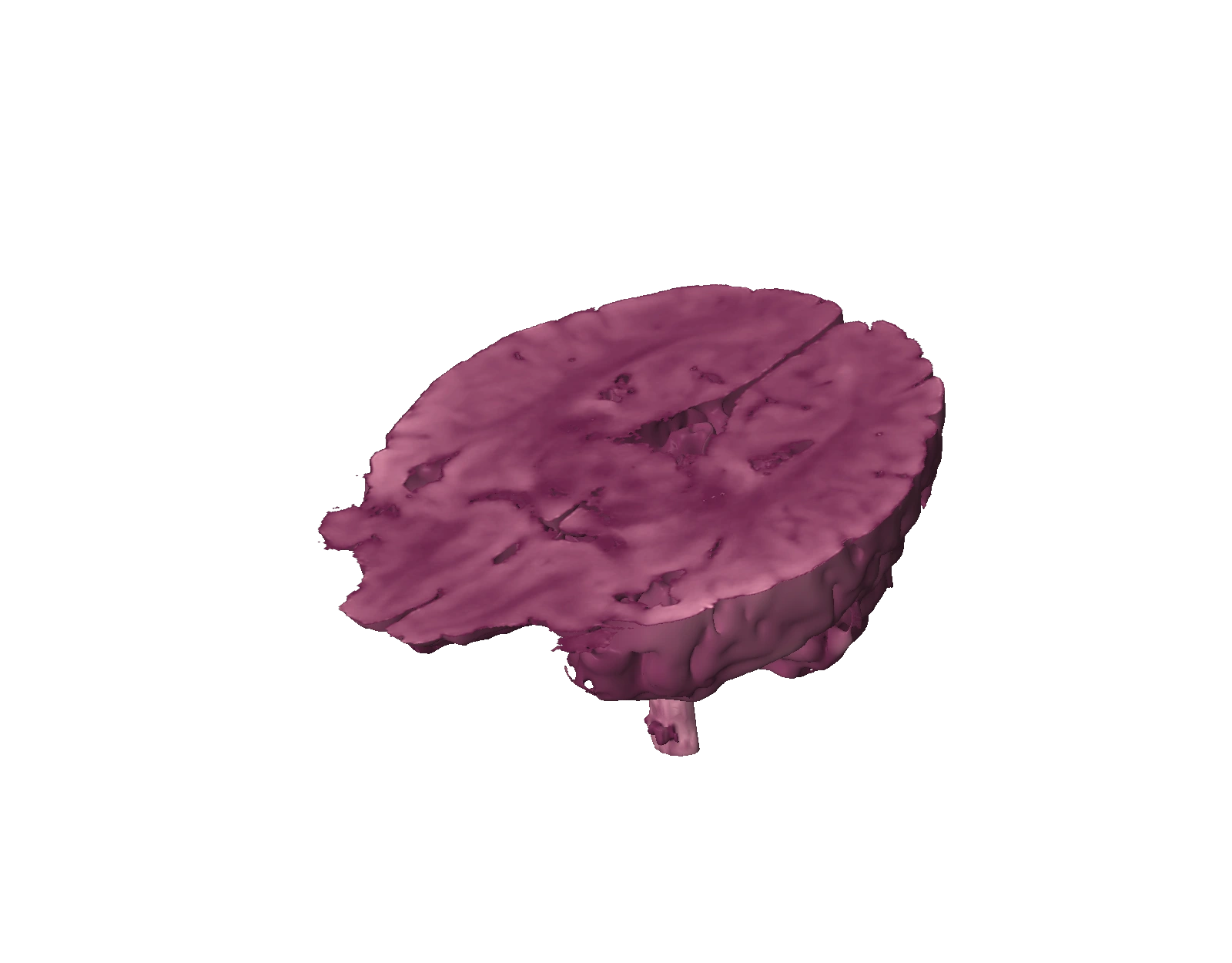
A bright outer shell could hide the character of the scan. I increased contrast, made the darkest regions transparent, and tied the exterior color to the image signal. The brain-only approximation also needed to keep the brain stem.
Smoothing took several passes. A surface could look cleaner while the coronal and sagittal cuts still showed stretched texture from the space between axial images. I separated those problems: refine the exterior, then smooth the cut-display signal while protecting the existing gap boundaries and brain-stem region.
Make exploration feel deliberate
Three draggable planes replaced separate slice panels. Blue marks axial cuts, red marks coronal cuts, and green marks sagittal cuts. They work together, so you can reveal a smaller region from more than one direction.
The outlines stay quiet until hovered. Four-way arrows mark the handles. The controls sit inside the viewer, and upright rotation limits, gentle snapping, and standard views help keep the model readable. The brain and planes rotate together under front lighting, so the side coming into view remains illuminated.
A project I can invite people into
The result is a rose-pink reconstruction that renders directly in the browser, with a smooth exterior and three cutting planes for exploring the interior.
The original experiment helped me engage with information that felt distant and difficult to access. Returning to it let me improve the object and the experience around it. The same curiosity is still there; now there is a clearer way for someone else to follow it.
About this case study +
The 2023–2024 history is documented in the original project account and Blender demonstration. The 2026 rebuild is based on the original DICOM files and the reconstruction and browser-interaction work completed for this release. Interpolated anatomy is approximate; this is a personal visualization project.